New treatment for brain swelling

Being treated for a brain tumor is scary enough, but if patients survive the surgery, radiation and chemo, they often have to take daily steroids to reduce swelling that can occur in the brain.These drugs have the potential to cause a number of unwanted side effects, but now there is a new option that is much safer.

if (self[‘plpm’] && plpm[‘Mid-Story Ad’]) document.write(‘

‘);if (self[‘plpm’] && plpm[‘Mid-Story Ad’]){ document.write(plpm[‘Mid-Story Ad’]);} else { if(self[‘plurp’] && plurp[’97’]){} else {document.write(”); } }if (self[‘plpm’] && plpm[‘Mid-Story Ad’]) document.write(‘

‘);When Paul Glover was diagnosed with a brain tumor, he never thought he would still be alive ten years later, and doing so much.

“What everybody told me is, ‘Yeah, life is over, pretty much,’” said Glover.

After surgery and chemo, Paul’s outlook was good, but the swelling in his brain took a toll on his body and caused neurological damage. Paul had to take steroids to fight it, but the side effects were harsh.

“… I didn’t feel like living. I couldn’t walk and half the time I would fall,” explained Glover.

Steroids caused him to gain nearly 80 pounds, and even forced the avid hunter into a wheelchair. The drugs can also cause bone loss, muscle weakness and anxiety.

So Paul was thrilled when he heard about a clinical trial that was testing a new therapy to replace steroids.

HCRF is the first agent in 40 years that was developed for brain tumor swelling.

“We are getting good, good results with some astonishing results in certain patients,” said Dr. Nicholas Avgeropoulus, from the Florida Hospital Cancer Institute.

When injected, HCRF works by stopping fluids in the brain tissue and reducing pressure in the skull. It even helps the body naturally produce its own steroids to reduce the swelling, without the side effects.

“The quality of life is such an important and understated factor in patients going through cancer treatment,” explained Avgeropoulus.

Something Paul knows all too well, now that he is off steroids and on the new drug.

Glover said, “Getting off steroids was wonderful. I cannot describe it … a big deal!”

Patients in the clinical trial inject the drug two times a day, and side effects may include redness.

Right now, researchers are enrolling patients for phase three of the clinical trial at about 35 centers around the country.

As reported in WNDU.om
New software to create an interactive 3-D map of the brain

As reported in Technology Review today.


medicad_x220.jpg

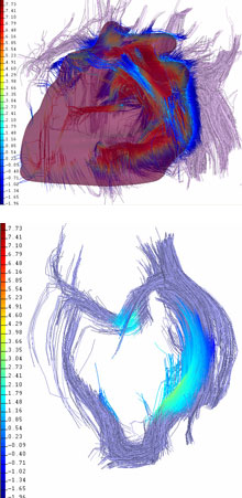
Researchers at Thomas Jefferson University Hospital, in Philadelphia, have developed software that integrates data from multiple imaging technologies to create an interactive 3-D map of the brain. The enhanced visualization gives neurosurgeons a much clearer picture of the spatial relationship of a patient’s brain structures than is possible with any single imaging methods. In doing so, it could serve as an advanced guide for surgical procedures, such as brain-tumor removal and epilepsy surgery.

The new imaging software collates data from different types of brain-imaging methods, including conventional magnetic resonance imaging (MRI), functional MRI (fMRI), and diffusion-tensor imaging (DTI). The MRI gives details on the anatomy, fMRI provides information on the activated areas of the brain, and DTI provides images of the network of nerve fibers connecting different brain areas. The fusion of these different images produces a 3-D display that surgeons can manipulate: they can navigate through the images at different orientations, virtually slice the brain in different sections, and zoom in on specific sections.

Currently, physicians typically view the images produced by MRI technologies individually, and they conceptually visualize what the images might look like combined. “Before this type of software package, I would put up an fMRI image and put up a regular MRI of the brain and try to match the two in my brain to try to get a 3-D sense of the right spot to make an incision,” says Ashwini Sharan, a neurosurgeon at the Jefferson Comprehensive Epilepsy Center.


Hope for the future

Chemical ‘Paint’ Helps Surgeons See Cancer’s Borders
07.16.07, 12:00 AM ET MONDAY, July 16 (HealthDay News) — Researchers say they’ve developed a tumor “paint” that illuminates cancerous cells and help surgeons spot the borders of tumors.A team at Seattle Children’s Hospital Research Institute and the Fred Hutchinson Cancer Research Center found that the paint — a protein derived from scorpions called chlorotoxin — helped surgeons distinguish between brain tumor cells and normal brain tissue during surgery.

“My greatest hope is that tumor paint will fundamentally improve cancer therapy. By allowing surgeons to see cancer that would be undetectable by other means, we can give our patients better outcomes,” study senior author Dr. James M. Olson said in a prepared statement.

The findings are in the July 15 issue of the journal Cancer Research.

Chlorotoxin is linked to a molecular “beacon” called Cy5.5. The use of chlorotoxin:Cy5.5 improves the likelihood that surgeons will be able to remove all cancerous cells during surgery without damaging surrounding healthy tissue, the researchers said. This is especially important for brain cancer patients. About 80 percent of malignant cancers recur at the edges of sites where tumors have been surgically removed.

Until now, there has been no way to allow surgeons to “see” tumors during surgery.

The researchers also noted that current technology, such as MRI, can distinguish tumors from healthy tissue only if more than one million cancer cells are present. Chlorotoxin:Cy5.5 is able to identify tumors with as few as 200 cancer cells, which means that it’s 500 times more sensitive than MRI.

The tumor paint has been successfully tested in mice, and pilot safety trials have been completed. The researchers are preparing required toxicity studies before they apply to the U.S. Food and Drug Administration for permission to begin human clinical trials with the tumor paint.